
Understand Your Knee Pain
Your knee is one complex joint, and if painful, its likely the cause of you being unable to walk, jump or run.
The key to getting better is understanding the structure of your knee so you can visualize what may be causing your pain.
Often its not just old age…
Knee Anatomy
The knee joint is compromised of the longest bone in the body, the femur (thigh bone), the tibia and fibula (shin bones) and the patella (knee cap). Along with these bones, there are 4 main ligaments (connecting bone to bone); the Medical collateral ligament (MCL), the lateral collateral ligament (LCL), the anterior cruciate ligament (ACL) and the posterior cruciate ligament (PCL). Moreover the knee joint contains many tendons (connecting muscle to bone) and menisci which are cartilaginous structures that help absorb shock in the knee and also have bursae which are fluid-filled sacs which help with the movement of the joint. The congruence of all of these structures is one of the main reasons the knee joint is one of the most complex joints in the body.
Bones of the knee joint:
There are four main bones which make up the knee joint;

Femur: The femur is the longest bone in the body and is a ball and socket bone. It runs from your hip to your knee.
Tibia: This bone helps make up the shin. It is the longer of the two bones within your shin. It is located on the inside of your leg and is connected to femur via the MCL.
Fibula: This is the other and shorter bone, which helps make up your shin. It is located on the outside of your knee and connects to the femur via the LCL.
Patella: The patella is also more commonly known as your knee cap. It is a triangular shaped bone which articulates with your femur and helps provide protection to the front of your knee.
Ligaments of the knee joint:

Ligaments are structures which help connect bone to bone. In doing so, they help give stability and help strengthen joints within your body.
Medical Collateral Ligament (MCL): Your MCL helps connect your femur (thigh bone) to your tibia(shin bone). This ligament is located on the inside of your knee. It helps provide your knee with support and helps prevent your knee from bending inward.
Lateral Collateral Ligament (LCL): Your LCL helps connect your femur( bone) to your fibula (shin bone). This ligament is located on the outside of your knee. It helps provide your knee with support and helps prevent your knee from bending outwards.
Anterior Cruciate Ligament (ACL): Your ACL joint is one of two ligaments which make up your cruciate complex, cruciate means cross and your ACL as well PCL cross to make this complex. The ACL in the front of your knee connects the femur to the tibia. It helps provide your knee with support and helps prevent your tibia from dislocating in front of your femur.
Posterior Cruciate ligament (PCL): Your PCL joint helps make up the cruciate complex. The PCL is located in the back of your knee joint and also connects the femur to the tibia. It helps provide your knee with support and helps prevent your tibia from dislocating behind your femur.
Meniscus:
Your meniscus are known as the shock absorbers of the knee (seen in the joint above). The meniscus are cartilaginous structures which are C shaped and help provide cushioning of the knee joint. They are located between the Femur and Tibia on both ends to help provide shock absorption. Each knee joint has two menisci, one that is located on the outside of the knee joint(lateral meniscus) and one the is located on the inside of the knee (medial meniscus).
Muscles:
The muscles of the knee joint can be grouped into three main categories; the anterior (front), the posterior (back), and medial (midline) compartments.
The anterior or front of the knee joint contains the tendons of your quadriceps muscles. The quadriceps are named as such because there are four (quad) part to this muscle. The four parts are the rectus femoris, vastus medialis, vastus lateralis, and vastus intermedius, together these muscles help extend the knee.
The posterior or back of your knee contains the tendons of the Hamstrings, calves and a couple of other smaller but important muscles for the knee. The hamstring muscles are made up of three smaller muscles; the semitendinosus, semimembranosus and the biceps femoris. These muscles work together to create the hamstrings and help bend your knee. Your gastrocnemius muscle is the part of your calves that connects to the knee and helps aid in bending of your knee as well as plantar flexing your foot.
Your Plantaris muscle also helps your bend your knee and helps your plantar flex your foot. Your popliteus muscle is another muscle located in the posterior part of the knee joint and helps with medial rotation and bending of the knee. Another muscle which helps provide the same type of movements is the Sartorius, although this muscles main functions involved movements in the hip.The muscle in the medial aspect of the knee is known as the gracilis and helps provide knee bending and medial rotation of the knee.

Knee injuries:
As we all know, the knee joint is one of the most used joints in the body. With this increased usage, there is always a risk of injuring the knee joint. If you’ve injured your knee and aren’t sure as to what could be causing it, we will list some of the most common knee injuries below;
Patellofemoral Pain Syndrome (PFPS):

PFPS is one of the most common injuries of the knee. It is also referred to as “runners knee” or “jumpers knee” due to the high incidence in runners and athletes involved in jumping sports. PFPS can be quite debilitating. In most cases of PFPS, the pain is a dull achy pain that is located in the front of the knee or around the knee cap. Sometimes severe forms of PFPS can also cause a sharp pain. The pain is usually exacerbated by activities such as climbing stairs, kneeling, and exercise. PFPS can be caused by many things; however, the main cause of this injury comes from intense athletic involvement, or it may stem from alignment issues of your knee cap. If you are having knee pain, it is always a great idea to see a healthcare provider to assess, diagnose, and help remedy the pain you are having.
Knee Osteoarthritis (Knee O.A):

Osteoarthritis is the most common type of arthritis in the world. It is caused by wear and tear on your joints. Your bones have cartilage that helps provide an ideal environment for your bones to slide and move, in essence, provide cushioning for the bones. Over time this wears away, and the result is O.A. Knee O.A is arthritis in the knee which occurs with time and use of the knee joint. The symptoms of knee O.A are swelling, sharp or achy pain, locking of the knee joint, and pain that is usually worst after a bout of rest. Knee O.A is usually found in people 50, and over, however, people who are younger can still be predisposed to the type of injury. The way to get an accurate diagnosis for this condition is to see your healthcare provider and speak to them about your symptoms.
Meniscal Tear:
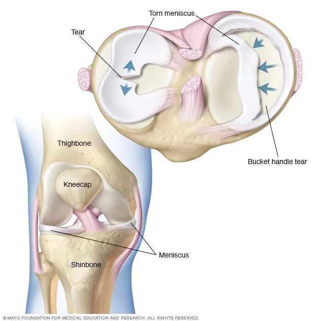
As mentioned earlier, your meniscus acts as the shock-absorber of the knee. If your knee is unable to adequately absorb the force put on it may cause you to injure the meniscus. The symptoms of a meniscus tear are a sharp pain either on the inside or outside of your knee, an inability to bend your knee, swelling and if it is very painful to put pressure on the affected side. You are more likely to injure this part of your knee if you are twisting your knee quickly while your foot is on the ground and your knee is bent. This type of motion usually occurs with those who are involved in sports or those who are lifting heavy weights and have their knees in the position mentioned above. Furthermore, because your menisci are cartilage, they also wear away with time, and you can potentially get a tear due to degenerative changes. It is always a good idea to see a healthcare professional if you are experiencing these symptoms.
ACL Injuries:

ACL injuries are most commonly found in athletes or people who have had direct trauma to the area. About 70% of ACL injuries are non-contact injuries; this means that the movements and force generated by the individual were the reason for the injury. An example of this would be someone who is running at full speed and now needs to stop very quickly and in doing so, cause themselves to get injured. Cutting movements and deceleration are the most common culprits for these non-contact ACL injuries. The other 30% of the ACL injuries involved some sort of direct contact or blow to the knee. If you have injured your ACL, a grading system will be used to assess severity. A grade 1 sprain is an injury to the ACL in which it has been damaged. However, it is still intact and is able to continue to stabilize the knee joint. A grade 2 sprain is an injury to the ACL in which there is a partial tear of the ligament; the knee joint usually will feel unstable and as though it is “giving out”. A grade 3 sprain is an injury to the ACL in which there is a full tear; there is a complete inability for this ligament to stabilize the knee joint. Always consult a healthcare provider if you have been feeling the above symptoms.
PCL Injuries:

PCL injuries are usually caused by direct impact on the ligament, unlike ACL injuries, which are a majority of non-contact injuries. PCL injuries are usually caused when there is a direct force applied to the knee when it is bent. These injuries usually occur in sports and in auto collisions when your knee strikes the dashboard. The symptoms of this injury are pain and a loss of stability in the knee joint. If you have injured your PCL, a grading system will be used to assess severity. A grade 1 sprain is an injury to the PCL in which it has been damaged. However, it is still intact and is able to continue to stabilize the knee joint. A grade 2 sprain is an injury to the PCL in which there is a partial tear of the ligament; the knee joint usually will feel unstable and as though it is “giving out”. A grade 3 sprain is an injury to the PCL in which there is a full tear; there is a complete inability for this ligament to stabilize the knee joint. Always consult a healthcare provider if you have are feeling the above symptoms.
MCL Injuries:

Your MCL helps stabilize the knee by preventing it from bending inwards. So any forces or movements which cause your knee to bend inwards beyond its normal limits can cause a sprain or even a tear of your MCL. For this reason, contact sports such as hockey and football tend to have increased rates of MCL injuries. If you are feeling pain, swelling, or tenderness on the inside of your knee, you may have injured your MCL. It is always a great idea to see a healthcare professional if you are experiencing any of these symptoms.
LCL Injuries:

Your MCL helps stabilize the knee by preventing it from bending outwards. So any forces or movements which cause your knee to bend outwards beyond its normal limits can cause a sprain or even a tear of your LCL. For this reason, LCL injuries are less prevalent than MCL injuries. With this injury, you may notice, pain, swelling, and tenderness on the outside of your knee joint. Be sure to check with a healthcare provider if you are feeling these symptoms.
Bakers Cyst
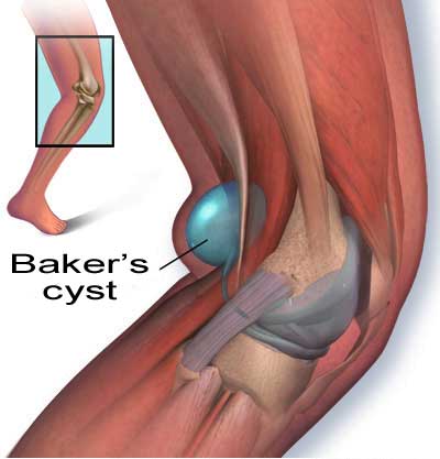
If you are noticing that there is increased swelling in the back of your knee this may in fact be a bakers cyst. The symptoms of this is noteable swelling and pain located in the back of your knee. This may even prevent you from being able to fully bend your knee. Bakers cysts have many causes and may be caused by; arthritis, gout, increased knee joint swelling, and a variety of the knee injuries mentioned above.
Diagnosis:
The key to getting the best results after a knee injury is to make sure you got and see your healthcare provider, so they are able to assess, diagnose and create a treatment plan to help you get back on your feet. The earlier you go to see your healthcare provider after an injury, the increased likelihood that you will be able to recover faster and get back to your pre-injury state.
Treatment:
There are many treatments for knee injuries depending on the type and severity of your injury. This is why it is so important to make sure that you get an accurate diagnosis and a comprehensive treatment plan sooner than later. Below we have listed some of the most common types of treatments for people with knee injuries;

Physical therapy: Getting physical therapy can be the major difference between getting you back on track and having long standing and persistent knee pain. For some types on knee pain, custom orthotics can provide significant relief. By addressing the reasons for your pain and creating a thorough and effective treatment protocol this can help get you on the road to recovery.
Pain medications: Your doctor may prescribe you anti-inflammatories, pain killers and or ointments to help your decrease inflammation and pain.
Cortisone injections: If you are having severe pain and physical therapy and pain medication are not providing relief your doctor may prescribe a cortisone injection.
Arthroscopic surgery: This is a medical procedure in which your surgeon is able to view, diagnose and treat your knee. Arthroscopic surgery is used for a number of injuries and is less invasive than open knee surgery and rehabilitation time is much faster when compared to open knee surgery as well.
Open Knee Surgery: In this surgery the knee is actually opened so that the surgeon can go in and replace or remove whatever is bothering the patient. This surgery is used in more severe cases and should always be discussed with a specialist before commencing.
Conclusion
The knee is complex and can have many different issues arise. Because the knee is a weight-bearing joint, it’s important to start managing them a soon as possible. With a better understanding of your knee pain, be sure to seek out quality help when needed and address your pains. For most of us, physiotherapy is enough to fix our pain and return our pain free lives. Understanding the source of your pain will be the first step on your journey to recovery.
Read Other Related Articles


